
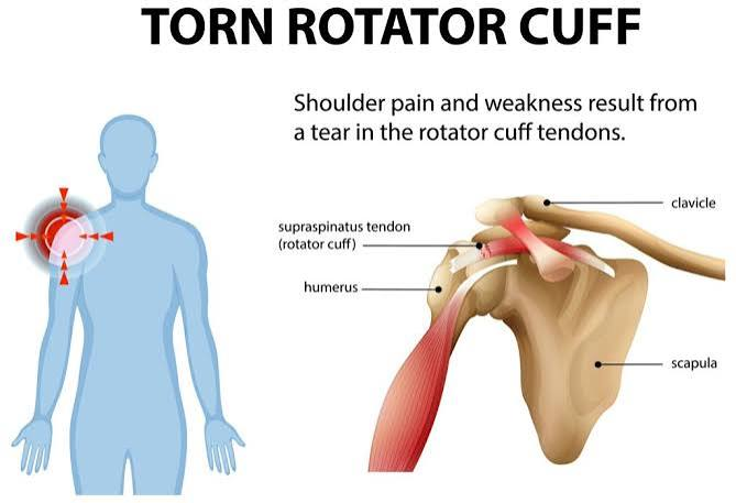
The shoulder is the most mobile joint in the body, and that mobility comes at a cost: stability depends heavily on a group of muscles and tendons called the rotator cuff, rather than on bone structure alone. When something in that system is irritated, torn, or simply overworked, the shoulder tends to announce it clearly, with pain reaching overhead, disrupted sleep on the affected side, and a weakness that makes reaching behind the back or lifting a bag feel unreliable.
Shoulder pain is also one of the areas where patients most often request imaging before any hands-on assessment has taken place, assuming a scan will reveal exactly what is wrong. Evidence indicates that this instinct, while understandable, often leads to confusion rather than clarity, since imaging findings in the shoulder are common in people without any pain at all. A structured clinical assessment usually comes first, and it usually tells a clinician most of what they need to know.
What the rotator cuff actually does
The rotator cuff is a group of four muscles and their tendons that wrap around the shoulder joint, holding the head of the upper arm bone securely in its shallow socket while allowing the wide range of motion the shoulder is known for. These tendons work constantly during everyday arm use, reaching, lifting, throwing, and carrying, which makes them prone to gradual wear as well as acute injury.
Rotator cuff problems exist on a spectrum, from tendon irritation and inflammation, through partial thickness tears, to full tears where the tendon has completely separated. Age plays a significant role. Degenerative changes in the rotator cuff become increasingly common through the fifties and beyond, often without the person ever noticing symptoms, which is central to why imaging alone can be misleading.
What a structured shoulder assessment involves
A thorough shoulder assessment starts with history: how the pain began, whether it followed a specific injury or developed gradually, what movements provoke it, and whether there is associated weakness, numbness, or night pain. This history alone often distinguishes between a likely rotator cuff issue, a joint capsule problem, and pain referred from the neck.
The physical exam includes testing the shoulder’s range of motion, strength in specific positions that isolate individual rotator cuff muscles, and a series of movements designed to reproduce or relieve the pain in characteristic patterns. A skilled clinician can often identify with reasonable confidence which structure is involved and how severe the problem likely is, using this hands-on assessment alone, before any imaging is considered.
Why shoulder imaging findings can mislead
One of the more counterintuitive findings in musculoskeletal research is how common rotator cuff changes are in people with no shoulder pain whatsoever. Studies scanning the shoulders of pain-free adults consistently find a meaningful proportion with partial tears, tendon degeneration, or other changes that would look concerning on a report, in people who have never had a symptom. This proportion increases substantially with age.
This does not mean imaging is worthless. It means that a scan result needs to be interpreted alongside the clinical picture rather than treated as an automatic verdict. A patient with a small tear visible on imaging and a clinical picture that matches good strength and function may not need the same treatment as a patient with the same imaging finding but significant weakness and functional limitation. The image is one piece of information, not the whole diagnosis.
When imaging actually changes the management plan
Imaging earns its place when the result is likely to change what happens next, rather than simply confirming what the clinical assessment already suggested. Several situations reasonably warrant it.
- Significant weakness alongside pain, which raises the possibility of a larger tear that might benefit from a different management pathway.
- A shoulder problem that has not improved after a genuine trial of appropriately dosed physiotherapy, where imaging helps clarify why.
- A history of significant trauma, such as a fall or a forceful injury, where a substantial structural injury is more likely.
- A case where the clinical picture is unclear or inconsistent, and imaging would help distinguish between competing possibilities.
Outside these situations, jumping straight to imaging before a trial of appropriate treatment often produces a report full of age-related findings that create worry without changing the plan a clinician would have recommended anyway.
What rehabilitation typically involves
For the large majority of rotator cuff and general shoulder pain presentations, a structured physiotherapy program is the first and most evidence-supported approach, including for many confirmed partial tears. The program typically progresses through phases: reducing pain and irritation, restoring range of motion, then progressively strengthening the rotator cuff and the surrounding shoulder blade muscles that support it.
Patients often want to protect a painful shoulder by avoiding movement, but evidence indicates that appropriately dosed loading, rather than rest, is what rebuilds the tendon’s capacity and resolves symptoms for most presentations. The skill lies in dosing the exercise correctly, enough to stimulate adaptation without provoking a flare, and progressing it as strength and tolerance improve over a period of weeks to a few months.
Consistency matters more than any single session. A short daily or near-daily routine of the prescribed exercises tends to outperform a longer but sporadic effort, since the tendon adapts to a steady pattern of appropriate load rather than an occasional intense one. Patients who track their symptoms and effort over the weeks of a program, rather than judging progress from any single day, tend to stay with it through the slower early stretches.
Managing sleep and daily activity during the recovery period
Shoulder pain has a well-earned reputation for disrupting sleep, particularly for patients who normally sleep on the affected side, and poor sleep can slow recovery by increasing overall pain sensitivity and reducing the body’s capacity to repair tissue. A pillow tucked under the affected arm for support, sleeping on the back or the unaffected side, and avoiding a fully flat position during an acute flare all reduce nighttime discomfort for many patients.
Daily activities often need temporary adjustment rather than complete avoidance. Reaching for items on high shelves, carrying heavy bags on the affected side, and overhead tasks like changing a lightbulb or reaching into a cupboard tend to provoke symptoms early in the recovery process. Modifying how a task is done, using the unaffected arm where possible or breaking a task into smaller movements, keeps a patient functional without forcing the shoulder into positions it is not ready for.
Ice or heat, used according to what a patient finds genuinely helpful rather than a rigid rule, can ease discomfort alongside the active rehabilitation program. None of these measures substitute for the structured exercise program that does the actual work of recovery, but they make the weeks of rehabilitation considerably more tolerable, which in turn supports consistency with the exercises that matter most.
Why coordinated assessment protects patients from unnecessary steps
A shoulder complaint that moves through a physician, a physiotherapist, and, when genuinely needed, imaging, works best when those pieces are connected. A physiotherapist’s detailed functional assessment can inform a physician’s decision about whether imaging is likely to add value, and a physician’s read of any imaging that is ordered can be interpreted alongside what the physiotherapist has already observed clinically, rather than in isolation.
This coordination reduces the number of patients who receive imaging that changes nothing, and it speeds up the path to appropriate treatment for those who do need it. Patients with shoulder pain, particularly pain that is persistent, worsening, or accompanied by significant weakness, should consult a qualified clinician, and those ready for a structured assessment can reach out to Calgary’s multidisciplinary care team to determine the right next step.
Assessment first, imaging when it earns its place
Shoulder and rotator cuff pain responds well to a structured approach: a thorough clinical assessment first, imaging reserved for situations where it will genuinely change the plan, and a progressive rehabilitation program for the great majority of cases. Jumping straight to a scan often produces more confusion than clarity, given how common incidental findings are.
Patients with shoulder pain should consult a qualified clinician for an individualized assessment rather than assuming imaging is the necessary first step. None of this replaces medical advice, but understanding the sequence, assessment, trial of treatment, imaging only when indicated, helps patients make sense of what a good shoulder workup should look like.
About the author: this article was contributed by the team at Primaris Health, a Calgary multidisciplinary clinic where physiotherapists and family physicians share one chart to assess and treat shoulder pain. The clinic builds structured rehabilitation plans first and coordinates imaging only when the clinical picture indicates it will change the course of treatment.

